

Case of the Week
- Sara Jiang, MD
- Jan 18, 2021
- 1 min read
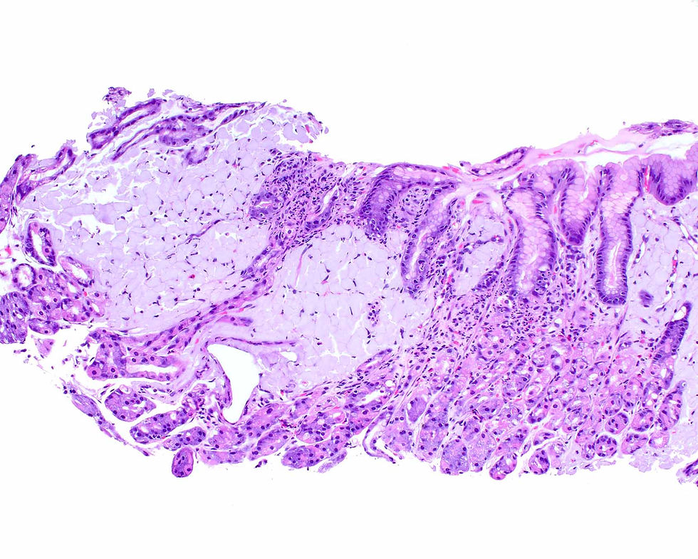
These images are from a fine needle aspiration biopsy of a thyroid nodule.
What is your diagnosis?


Answer: Medullary thyroid carcinoma. Comment: The Diff-quik image (A) shows a cellular sample of monomorphic cells with abundant cytoplasm and eccentrically placed nuclei. Many isolated cells are present. The alcohol-fixed smear (B) shows a dishesive population of cells with abundant cytoplasm and scattered enlarged nuclei. Amorphous waxy material suggestive of amyloid is present in the upper right side of the field.
This case was provided by Dr. Xiaoyin “Sara” Jiang, MD, is Associate Professor of Pathology at Duke University and Director of the Head and Neck Service. Her book Survival Guide to Cytopathology published by Innovative Science Press is now being reviewed and should be released in the Summer.


Comments