

Case
- Andres Matoso, MD
- Jul 26, 2021
- 1 min read
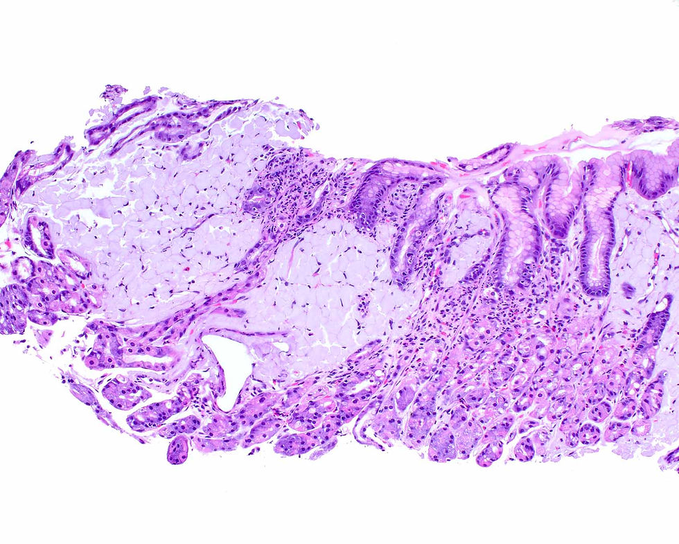
Normal colonic mucosa can mimic prostate adenocarcinoma morphologically with rows of small glands with cells with large nuclei, prominent nucleoli, and amphophilic cytoplasm. Note that they lack goblet cells due to procedural compression. These glands stain like prostate cancer with a triple stain (PIN4), showing diffuse staining for arginase (red) and lack of staining for p63 and HMWCK. The key to avoid this morphological and immunohistochemical pitfall is to identify the lamina propria (arrow) surrounding the colonic glands. Additionally, positive CDX2 immunostaining confirms the intestinal origin of the glands.

This case was provided by Dr. Andres Matoso, Associate Professor of Pathology, Urology and Oncology, and the Director of the Immunohistochemistry Laboratory at Johns Hopkins Hospital. The book Survival Guide to Prostate Pathology that he and Dr. Jonathan Epstein wrote was recently released. You can order your copy at:
info@innovativepathologypress.com
1-703-350-4308/703-340-3198


Comments